Home / Healthcare Professional / Gaucher’s Disease

Diseases

Click here to view more Diseases

Gaucher Disease

Gaucher disease (GD) is a sphingolipidoses, a subgroup of lysosomal storage diseases. It is the most common lysosomal storage disease, having an autosomal recessive inheritance and resulting from a deficiency of the glucocerebrosidase enzyme due to a genetic defect on the glucosylceramidase beta (GBA1) gene. The enzyme deficiency results in the inability to break down glucosylceramide in lysosomes, and lipid-laden macrophages accumulate in the tissues.

Gaucher disease (GD) is the commonest lysosomal storage disorder (LSD) with an estimated global incidence of 1: 40,000 to 1:60,000 live births. In the Ashkenazi (Eastern European) Jewish population, with a carrier frequency of 6% compared to 0.7% to 0.8% of the non-Jewish population. Gaucher disease's exact prevalence in India is not fully documented due to a lack of centralized registries, due to the tradition of consanguineous marriages in parts of the country, it seems likely that the frequency of Gaucher disease may be higher in India. The most common form of Gaucher disease is type 1, which has a very variable phenotype ranging from early childhood symptoms to no symptoms throughout life but typically does not have a neurological component. In contrast to type 1, types 2 and 3 are rare and affect the central nervous system.

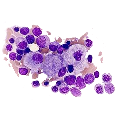
Glucosylceramide, the accumulated glycolipid, is primarily derived from the phagocytosis and degradation of senescent leukocytes and erythrocyte membranes. The glycolipid storage gives rise to the characteristic Gaucher cells, macrophages engorged with lipid with a crumpled–tissue-paper appearance and displaced nuclei. The factors that contribute to neurologic involvement in patients with types 2 and 3 disease are still unknown but may be related to the accumulation of a cytotoxic glycolipid, glucosylsphingosine, in the brain due to the severe deficiency of glucocerebrosidase activity or to neuroinflammation. Glucosylceramide accumulation in the bone marrow, liver, spleen, lungs, and other organs contributes to pancytopenia, massive hepatosplenomegaly, and, at times, diffuse infiltrative pulmonary disease. Progressive infiltration of Gaucher cells in the bone marrow may lead to thinning of the cortex, pathologic fractures, bone pain, bony infarcts, and osteopenia.

Individuals with Gaucher disease may exhibit the following signs and symptoms:

Enlargement of the spleen (splenomegaly)
  • Often one of the earliest signs occurring in childhood or adolescence. An enlarged spleen can lead to hypersplenism, where the spleen destroys blood cells, worsening anemia, thrombocytopenia and leukopenia.
  • Splenic Infarcts though rare, may occur and cause abdominal pain and digestive issues.
Enlargement of the liver (hepatomegaly)
  • May manifest in childhood or adolescence accompanying an enlarged spleen, and may result in liver dysfunction, though it is less common than splenomegaly
Bone Abnormalities
  • Includes bone pain, fractures and skeletal abnormalities, which can occur at any age. Individuals with Gaucher disease who experience symptoms in childhood may develop widening of the long bones, known as ‘Erlenmeyer flask deformities.’ Others may develop abnormal bone density or other skeletal changes later in life which can lead to bone fragility and fractures.
  • Episodes of severe bone pain called “bone crises” due to infarctions or the death of bone tissue may occur.
  • Osteonecrosis is the most severe bone complication, which can lead to significant bone deformities, often requiring pain management and/or surgery.
Hematological abnormalities
  • Anemia and thrombocytopenia can occur at various stages and may worsen over time.
    Anemia or low red blood cell count can lead to fatigue and weakness.
  • Low platelet counts can increase the risk of bruising and bleeding.
Neurological complications (in GD2 and GD3)
  • Includes swallowing difficulties, seizures, muscle stiffness, coordination problems and developmental delay, typically appearing in infancy or childhood. The most common indicator of brain involvement in GD is abnormally slowed eye movements or inability to move the eyes horizontally

The diagnosis of Gaucher disease depends upon finding a low GBA1 enzyme level in peripheral blood leukocytes and establishing the presence of mutant alleles in the GBA1 gene. Even though only a blood sample is needed to diagnose Gaucher disease, some patients undergo unnecessary invasive bone marrow or liver biopsy before making a correct diagnosis


  • Laboratory Studies: CBC Count, Liver Function Enzyme Testing and Coagulation
  • Enzyme Activity : Diagnosis is confirmed through measurement of glucocerebrosidase activity in peripheral blood leukocytes. Less than 15% of mean normal activity is diagnostic for Gaucher disease
  • Genotype Testing: In some ethnicities, sequencing of the exons of GBA1 may be necessary to establish the genotype
  • Imaging
  • Ultrasonography may reveal abdominal organomegaly
  • MRI may be useful in revealing early skeletal involvement (avascular necrosis, spinal degradation, and degree of bone marrow infiltration).
  • Radiography may reveal skeletal manifestations and pulmonary involvement.
Bone Marrow Aspiration
  • In the past, the diagnosis was made by finding classic glycolipid-laden macrophages in bone marrow aspirate
Liver Biopsy
  • A liver biopsy may be performed to evaluate unexplained hepatomegaly
 

Management

  • Treatment for Gaucher disease falls into 2 categories: enzyme replacement therapy and substrate reduction therapy.
  • The FDA has approved imiglucerase and velaglucerase alfa for Gaucher disease types 1 and 3 enzyme replacement therapy. Enzyme replacement therapy typically cannot replace an enzyme deficient in the brain due to the blood-brain barrier and, therefore, is not effective for treating the central nervous system problems associated with types 2 and 3 Gaucher disease. Enzyme replacement therapy will help with the "non-brain" signs and symptoms associated with type 3 Gaucher disease, eg, enlarged organs and skeletal issues. Enzyme replacement therapy does not correct the underlying genetic defect and only relieves signs, symptoms, and ongoing damage caused by the accumulation of toxins.
  • Substrate reduction therapy is an orally administered small-molecule drug (not protein) that relies on a strategy distinct from enzyme replacement therapy. In substrate reduction therapy, the goal is to reduce substrate levels such that toxic accumulation of the substrate's subsequent degradative product is diminished to a clinically less toxic level.
  • In the case of Gaucher disease, the goal is to use substrate reduction therapies to inhibit the first committed step in glycosphingolipid biosynthesis. There are 2 FDA-approved substrate reduction therapy drugs to treat patients with Gaucher disease: eliglustat and miglustat. Eliglustat, a glucosylceramide synthase inhibitor, is indicated only for type 1 Gaucher disease and does not effectively cross the blood-brain barrier. It is not yet known if eliglustat is safe or effective in children. Miglustat can cross the blood-brain barrier and potentially benefit types 2 and 3 Gaucher disease.
 

Eliglustat

  • Eliglustat is a glucosylceramide synthase inhibitor. It was approved in August 2014 as first-line treatment for the long-term treatment of adults with Gaucher disease type 1. The dose of eliglustat is determined by establishing the patient’s CYP2D6 phenotype (ie, extensive metabolizers [EM], intermediate metabolizers [IM], or poor metabolizers [PM]).
 
Indication Dose Contraindications Adverse Effects
Eliglustat is a glucosylceramide synthase inhibitor indicated for the long-term treatment of adult patients with Gaucher disease type 1 who are CYP2D6 extensive metabolizers (EMs), intermediate metabolizers(IMs), or poor metabolizers (PMs) as detected by an FDA-cleared test. EMs and IMs: 84 mg orally twice daily. PMs: 84 mg orally once daily. Taking a strong or moderate CYP2D6 inhibitor concomitantly with a strong or moderate CYP3A inhibitor. Most common adverse reactions (≥10%) are: fatigue, headache, nausea, diarrhea, back pain, pain in extremities, and upper abdominal pain.
 

New Innovations

New innovations in Gaucher disease treatment include gene therapy, brain-targeted therapies, and small molecule chaperones, offering potential for long-term benefits and reducing the need for continuous enzyme replacement therapy. Additionally, research focuses on improving current enzyme replacement therapies (ERT) with technologies like nanoERT and exploring new delivery methods like intranasal routes for brain delivery.


Challenges and Future Directions

Gaucher disease, a lysosomal storage disorder, presents significant challenges in diagnosis, treatment, and long-term management, particularly for the more severe forms. While enzyme replacement therapy (ERT) has revolutionized the management of Type 1 Gaucher disease, challenges remain with its cost, delivery limitations, and potential for antibody development. Future directions focus on developing targeted therapies, addressing the neurological complications of Type 3, and ensuring equitable access to treatment, especially in resource-poor countries.

References

  • Dursun, H., Yildizhan, E, et al. Overall assessment of patients with type 1 Gaucher disease: a single-centre’s experience. J Rare Dis 2, 14 (2023).
  • Wang M, Li F, Zhang J, et al. Global Epidemiology of Gaucher Disease: an Updated Systematic Review and Meta-analysis. J Pediatr Hematol Oncol. 2023 May 01;45(4):181-188
  • Ratna Dua Puri1, Seema Kapoor, et al. Diagnosis and Management of Gaucher Disease in India Consensus Guidelines of the Gaucher Disease Task Force of the Society for Indian Academy of Medical Genetics and the Indian Academy of Pediatrics. Indian Pediatr 2018;55:143-153
  • William L. Stone; Hajira Basit, et al. Gaucher Disease. StatPearls. Last Update: November 12, 2023.
  • https://rarediseases.org/rare-diseases/gaucher-disease/#symptoms
  • Stirnemann J, Belmatoug N, et al.A Review of Gaucher Disease Pathophysiology, Clinical Presentation and Treatments. Int J Mol Sci. 2017 Feb 17;18(2)
  • https://www.accessdata.fda.gov/drugsatfda_docs/label/2014/205494Orig1s000lbl.pdf
  • Richard Sam 1, Emory Ryan, et al. Current and emerging pharmacotherapy for Gaucher disease in pediatric populations. Expert Opin Pharmacother. 2021 Mar 25;22(11):1489–1503.
  • Vijay Bohra and Velu Nair . Gaucher's disease. Indian J Endocrinol Metab. 2011 Jul-Sep;15(3):182–186
  • Pramod K Mistry , Grisel Lopez et al. Gaucher Disease: Progress and Ongoing Challenges. Mol Genet Metab. 2016 Nov 17;120(1-2):8–21.