Diseases
Click here to view more Diseases
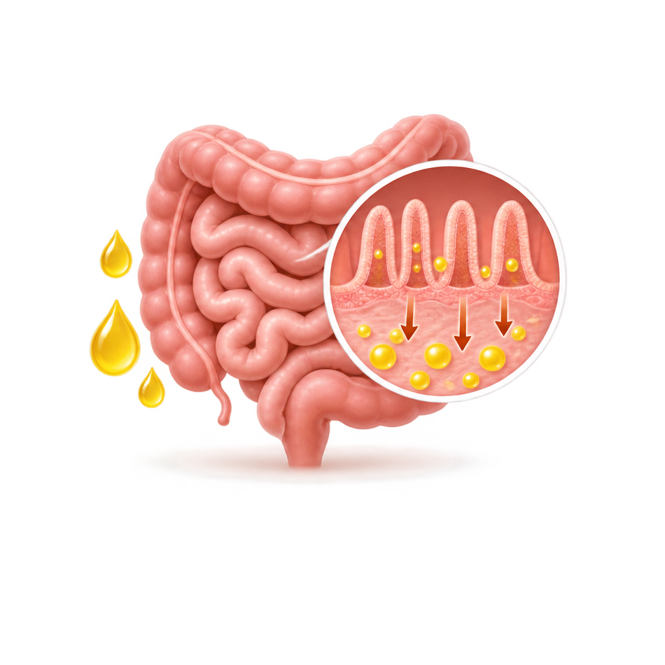

Fat malabsorption is the inadequate digestion or absorption of dietary lipids within the gastrointestinal tract. Because normal lipid absorption requires coordinated activity among pancreatic enzymes, bile acids, the intestinal mucosa, and the lymphatic system, dysfunction in any of these components impairs dietary fat utilisation.
Physiologically, gastric mixing disperses dietary fat, while pancreatic lipase hydrolyses triglycerides in the small intestine. Bile acids emulsify these lipids into mixed micelles (containing fatty acids, monoglycerides, cholesterol, and fat-soluble vitamins) for uptake by enterocytes. Within the enterocytes, lipids are reassembled into chylomicrons before entering lymphatic circulation. Disruption at any stage reduces lipid absorption and secondary uptake of fat-soluble vitamins (A, D, E, and K).
The clinical impact of fat malabsorption depends on the duration, severity, and primary etiology. Chronic malabsorption leads to inadequate lipid absorption, steatorrhoea, nutrient deficiencies, and progressive weight loss. In paediatric populations, the resulting nutritional insufficiency can impair growth and development, making routine nutritional assessment essential.
| Gastrointestinal Features | Nutritional Features |
|---|---|
|
|
The management of fat malabsorption requires a dual-targeted framework focused on resolving the primary etiologic pathology and repleting systemic nutritional deficits. Treatment must be precision-tailored to the specific site of digestive breakdown.
Cholestasis is a disorder characterised by the impaired formation, secretion, or flow of bile, leading to the retention of biliary constituents within the liver and their accumulation in systemic circulation. It arises from hepatocyte dysfunction, defective bile transport molecular mechanisms, or physical obstruction within the intrahepatic or extrahepatic biliary system.
Clinically significant cholestasis disrupts intestinal micelle formation by reducing luminal bile acid delivery, impairing the digestion and absorption of dietary lipids and fat-soluble vitamins (A, D, E, and K). In infants, early recognition of conjugated hyperbilirubinaemia is vital to differentiate it from physiological jaundice and to initiate timely, life-saving interventions.
Neonatal cholestasis is defined by an elevation of conjugated or direct bilirubin during the neonatal period. Persistent infantile jaundice must not be assumed to be physiological or prolonged unconjugated hyperbilirubinaemia.
Cholestatic disorders are classified based on the anatomical site and mechanism of impaired bile flow:
| Category | Pathophysiologic Mechanism | Key Conditions & Etiologies |
|---|---|---|
| Extrahepatic Cholestasis | Mechanestasis Intrahepaticical obstruction or anatomical abnormalities involving the extrahepatic biliary tree, preventing normal bile drainage. |
|
| Intrahepatic Cholestasis | Defective hepatocellular function, abnormal intrahepatic bile ducts, or mutations in canalicular transport proteins. |
|
Normal bile flow relies on hepatocyte transporter proteins secreting bile acids into the canalicular space, followed passively by water and electrolytes. Genetic mutations or acquired defects in these transporters cause intracellular retention of bile components. Intrehepatic accumulation of bile acids triggers a detergent-like cytotoxic effect on biological membranes, driving hepatocellular injury, localized inflammation, and progressive fibrotic liver disease. Physical mechanical obstructions cause similar upstream cellular damage while simultaneously starving the intestine of bile.
Because chronic cholestasis impairs lipid and micronutrient absorption, serial nutritional assessments are an essential component of long-term medical management. Clinicians should systematically monitor:
Persistent cholestasis prevents normal micellar incorporation, placing patients at high risk for clinical micronutrient deficiencies:
Cystic fibrosis (CF) is an autosomal recessive, multisystem disorder caused by pathogenic variants in the CFTR gene on chromosome 7, which encodes an epithelial chloride and bicarbonate channel. Impaired CFTR function disrupts trans-epithelial ion and water transport, producing hyperviscous secretions that cause ductal obstruction, chronic inflammation, and tissue remodelling across respiratory, gastrointestinal, pancreatic, and hepatobiliary systems. Advances in new-born screening, nutritional protocols, and CFTR modulator therapies have substantially altered disease progression and clinical outcomes.
The failure of apical chloride and bicarbonate transport induces mucosal dehydration and macromolecular protein precipitation across targeted abdominal organs:
Inadequate intraluminal release of pancreatic lipase prevents the hydrolysis of dietary triglycerides into absorbable fatty acids and monoglycerides. The resulting lipid malabsorption drives systemic caloric deficits and compromises the absorption of fat-soluble vitamins (A, D, E, and K).
| System | Key Clinical Signs & Manifestations |
|---|---|
| Gastrointestinal & Pancreatic |
|
| Hepatobiliary |
|
| Micronutrient Deficiencies |
|
CF diagnosis relies on a combination of characteristic clinical features, screening findings, and documented CFTR channel dysfunction:
Exocrine pancreatic insufficiency (EPI) is a physiological state characterised by the deficient secretion or inadequate luminal delivery of pancreatic digestive enzymes and bicarbonate. This deficiency severely compromises macronutrient hydrolysis within the small intestine.
Because pancreatic lipase is uniquely sensitive to parenchymal dysfunction and luminal acidification, lipid maldigestion is typically the earliest and most severe consequence of EPI. Unchecked fat malabsorption drives debilitating gastrointestinal symptoms, progressive weight loss, and secondary deficiencies of fat-soluble vitamins (A, D, E, and K).
EPI arises from progressive destruction of functional pancreatic parenchyma, mechanical obstruction of the main pancreatic duct, or postsurgical disruptions of gastroduodenal synchronization.
The clinical presentation reflects the extent of exocrine decline, the compensatory capacity of gastric/lingual lipases, and the underlying primary pathology.
| Gastrointestinal Features | Systemic & Nutritional Complications |
|---|---|
|
|
Diagnostic evaluation is indicated for patients presenting with classic gastrointestinal symptoms, unexplained weight loss, or an established high-risk predisposing condition.
The 13C-mixed triglyceride breath test evaluates fat digestion by pancreatic lipase. Following ingestion of a test meal containing a ^13C-labelled triglyceride, digestion generates ^13CO₂, which is subsequently measured in exhaled breath.
The therapeutic framework for EPI targets the normalization of nutrient digestion, weight maintenance, and prevention of metabolic bone or micronutrient diseases.
Celiac disease is a chronic, systemic immune-mediated enteropathy triggered by dietary gluten ingestion (proteins found in wheat, rye, and barley) in genetically susceptible individuals. The resulting autoimmune mucosal inflammation causes blunting of the small-intestinal microvasculature and villous architecture, diminishing absorptive surface area and inducing widespread malabsorption of macronutrients, minerals, and fat-soluble vitamins (A, D, E, and K).
Celiac disease develops through an interplay between dietary gluten exposure, genetic susceptibility, and coordinated innate and adaptive immune responses.
Because the proximal small intestine is the primary site of nutrient uptake, extensive enterocyte destruction compromises multiple metabolic pathways:
| Gastrointestinal Features | Extra-Intestinal & Nutritional Manifestations |
|---|---|
|
|
Serves primarily for its high negative predictive value (NPV). The absence of both HLA-DQ2 and HLA-DQ8 makes celiac disease virtually impossible, proving useful to exclude the diagnosis in ambiguous cases or patients already on a gluten-free diet.
Small intestinal bacterial overgrowth (SIBO) is a clinical syndrome characterised by an abnormal quantitative increase and/or taxonomic shift in the microbial population within the small bowel. Unlike the colon, which houses a dense microbial ecosystem, the small intestine maintains a low bacterial burden through an array of physiological clearative and antimicrobial barriers.
When these homeostatic defenses are compromised, proliferating bacteria ferment luminal carbohydrates and deconjugate bile salts, driving prominent gastrointestinal distress and, in severe or protracted cases, structural enterocyte irritation with subsequent malabsorption of macronutrients and fat-soluble vitamins (A, D, E, and K).
The restriction of microbes to the large intestine depends on the coordinated execution of multiple defense mechanisms. SIBO develops secondarily when one or more of these barriers fail:
Intestinal Peristalsis & the Migrating Motor Complex (MMC): Functions as an electrochemical "housekeeper," mechanically sweeping residual contents and bacteria downstream into the colon during fasting phases.
Proliferating bacteria compete with the host for nutrients and fundamentally alter the intraluminal environment. Intestinal microbes prematurely deconjugate and dehydroxylate bile acids, rendering them incapable of forming the mixed micelles necessary for lipid transport. This specific breakdown step causes progressive fat malabsorption and steatorrhoea. Additionally, bacteria consume luminal vitamin B₁₂ (cobalamin), potentially precipitating a macrocytic anemia despite adequate dietary intake.
| Gastrointestinal Features | Nutritional & Extra-Intestinal Features |
|---|---|
|
|
Because clinical manifestations overlap considerably with other functional and structural bowel disorders, objective diagnostic confirmation is required.
Therapy must simultaneously eradicate the overgrowth, manage symptoms, address the root cause, and correct metabolic deficits.
Disclaimer: For healthcare professional use only. The clinical information regarding fat malabsorption on this page is for peer educational purposes and does not substitute for independent professional medical judgment. Clinicians must verify all diagnostic protocols and treatment plans against current medical standards and individual patient presentations.

Disclaimer

 Healthcare
Professional